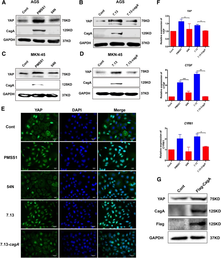
Fig. 5.
a, b AGS cells were infected with all H. pylori strains at MOI of 200 for 6 h. Levels of YAP and CagA were assessed by Western blotting in AGS cells co-cultured with PMSS1 and its ΔcagA mutant 54 N (a) or 7.13 and its CagA− mutant (b). c, d YAP and CagA levels were detected in MKN-45 cells. e Expression and localization of YAP (green) visualized by immunofluorescence. The blue-fluorescent DAPI was used for nuclear staining. f mRNA levels of YAP and YAP downstream target genes were assessed by qRT-PCR. Data for gene expression were the mean ± SEM of 3 independent experiments. g Representative Western blot for CagA and YAP in AGS cells transfected with the recombinant CagA protein

