
Figure 1.
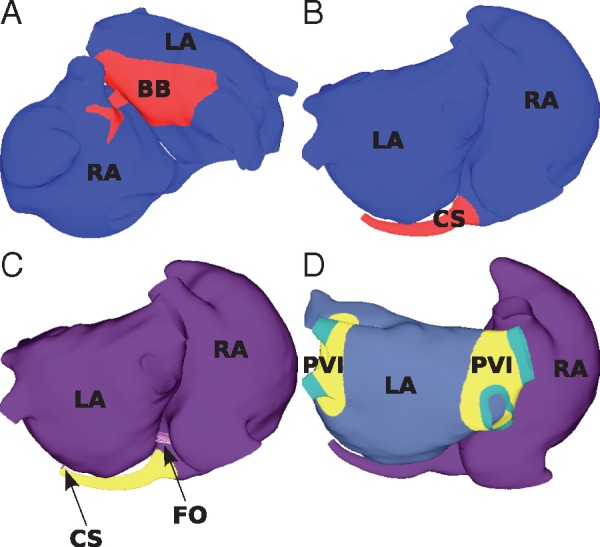
Interatrial connections. The LA and RA models are connected at (A) BB; (B) the CS; and (C) the FO. (D) To isolate the PVs, the tissue shown in yellow (labelled PVI) is set to non-conductive. The interatrial structures are connected to the LA and RA using discrete resistance line elements, shown as purple lines and indicated by the arrows in (C). BB, Bachmann’s bundle; CS, coronary sinus; FO, fossa ovalis; LA, left atrium; PVs, pulmonary veins; PVI, pulmonary vein isolation; RA, right atrium.