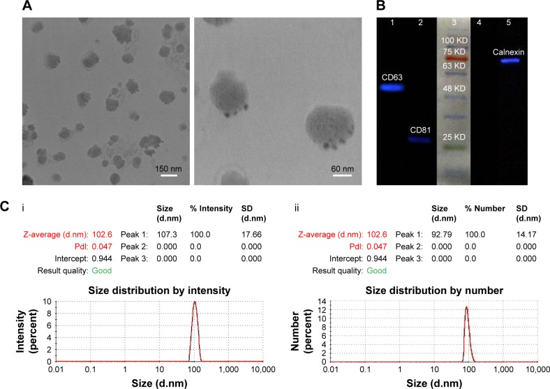
Figure 2.
Characterization of exosomal particles.
Notes: (A) Transmission electron micrograph of negatively stained exosomes with a diameter of 30–150 nm, labeled for CD63 (Low magnification, 13,000×, scale bar =150 nm, and high magnification, 40,000×, scale bar =60 nm). (B) Western blot analysis of positive and negative exosomal CD markers in both isolated exosomes and cellular lysate: (1) CD63 expression in exosomes lysate, (2) CD81 expression in exosomes lysate, (3) protein marker, (4) non-expression of calnexin in exosomes lysate, (5) the existence of calnexin in the cell lysate. (C) Size distribution by intensity (i) and size distribution by number of MSCs-Exo (ii).
Abbreviations: CD, cluster of differentiation; MSCs-Exo, mesenchymal stem cells-derived exosomes; Pdi, polydispersity index.

