Figure 8.
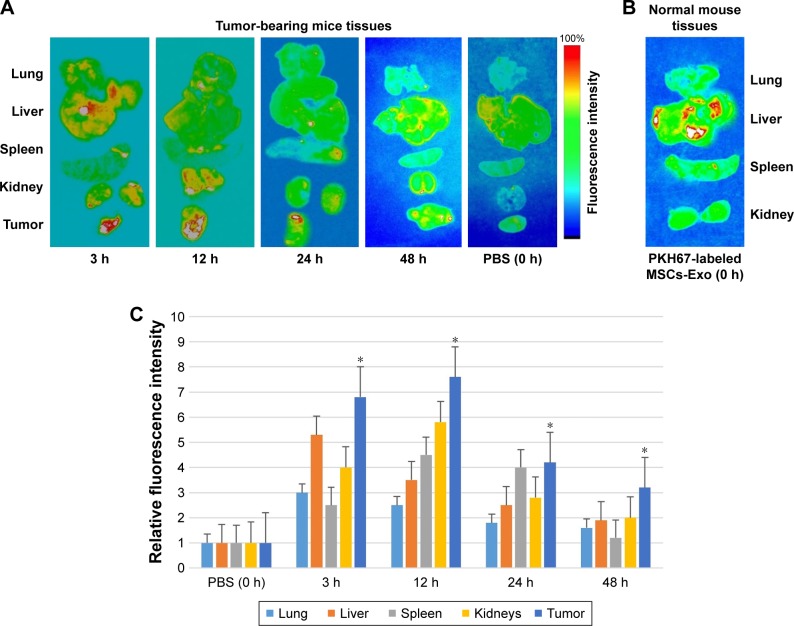
In vivo tracking of fluorescently labeled MSCs-derived exosomes in harvested tissues from tumor-bearing or control mice.
Notes: (A) Exosomes labeled with PKH67 were intravenously injected (30 µg of purified exosomes from MSCs) into mice bearing TUBO tumors. Lung, liver, spleen, kidney, and tumor tissues were harvested after 3, 12, 24, and 48 hours postinjection for ex vivo imaging. The fluorescent signal of PKH67-labeled MSCs-Exo was detected using IVIS. (B) The lung, liver, spleen, and kidney were also harvested from normal mice injected with 30 µg of labeled MSCs-Exo as control immediately after injection and fluorescent signal was acquired using IVIS. (C) The relative mean fluorescence intensity in the lung, liver, spleen, kidneys, and tumor tissues as a function of time after intravenous injection of fluorescently labeled MSCs-Exo. The fluorescence intensity in tumor tissues dissected from the mice injected with fluorescent exosomes was significantly higher compared to the fluorescence intensity in tumor tissue dissected from the control mouse (*P<0.05).
Abbreviations: IVIS, in vivo imaging system; MSCs-Exo, mesenchymal stem cells-derived exosomes.

