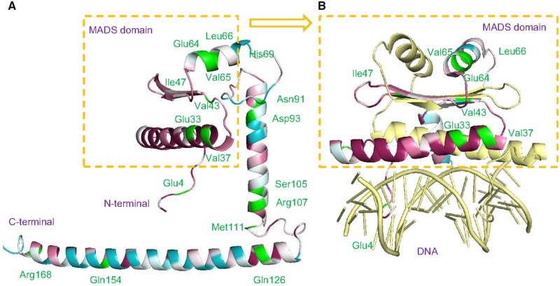
Fig. 6.
—Positive selection sites in the three-dimensional structure of the SVP protein. Residues are marked with different colors according to the degree of conservation. Conserved sites, magenta; variable sites, blue; and positive selection sites, green. (A) Positive selection sites shown in a variation of the three-dimensional structure of the SVP protein. (B) The MADS-box domain binding to DNA.

