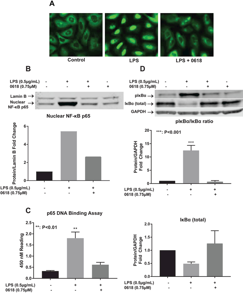
Figure 4:

CYD0618 suppresses classic NF-kB pathway LX-2 cells treated with LPS, CYD0618, or both CYD0618 and LPS for 1 hour underwent (A) Immunofluorescence staining for p65, (B) nuclear fraction Western blot with antibodies for NF-κB p65, (C) p65 DNA binding ELISA assay with 10 μg of nuclear protein, and (D) whole cell lysate Western blot with antibodies for NF-κB inhibitory protein IκBα and phosphorylated-IκBα. P-values shown compared to vehicle. Densitometric analyses of bands were quantified and data expressed as fold of control normalized to GAPDH for cytosolic protein or Lamin A for nuclear protein.
