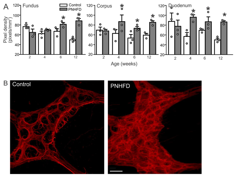
Figure 6. PNHFD increased glial cell density in the myenteric plexus.
A. Increase in GFAP-IR pixel cell density in myenteric ganglia of the fundus (left), corpus (middle), and duodenum (right). This increase was observed by 4 weeks of age, and maintained throughout the duration of the study (N=3 per data point; *P<0.05; one way ANOVA followed by post hoc Bonferroni comparison between groups)
B. Representative images of the myenteric ganglia in the corpus of 6 week old rats showing increased glial cell density in a PNHFD (right) vs control (left) myenteric ganglion. Scale bar = 20μm

