FIGURE 3.
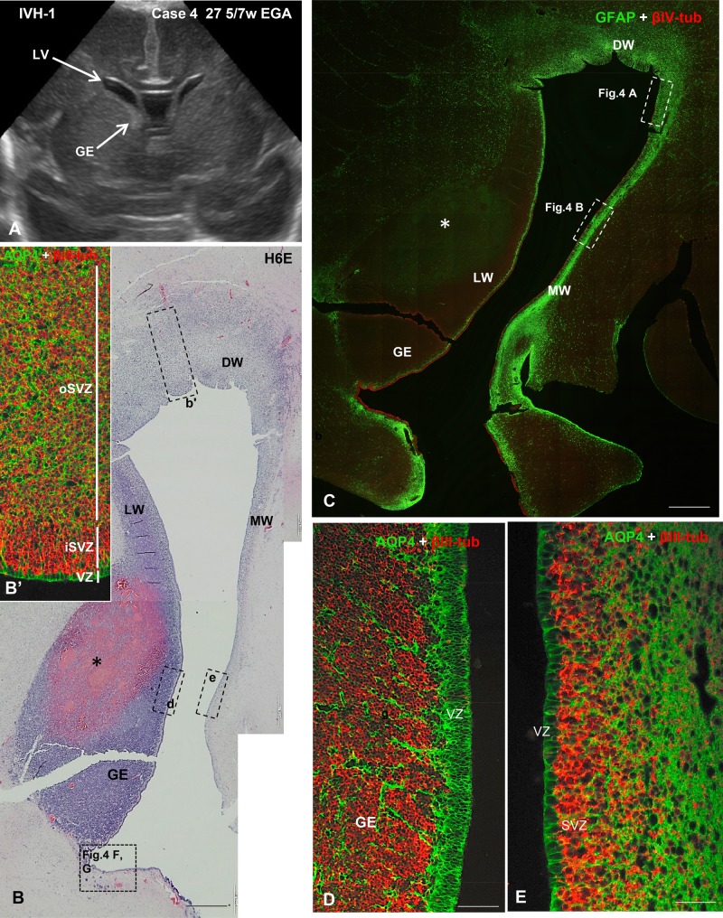
US image and photomicrographs from Case 4 illustrating the cytology and cytoarchitecture of the VZ typically found in IVH-1 cases. (A) A coronal US taken 2 days before death showing relatively small LVs and a prominent GE in the lateral wall of the frontal horn. (B) A H&E-stained section through the LW, MW, and dorsal (DW) walls of the LV showing a large hemorrhage (asterisk) in the GE. Dashed-line boxes demarcate regions shown in B’ (b’), D (d) and E (e) of this figure, as well as in Figure 4F, G. (B’) Area similar to that framed in b’, immunostained for AQP4 and βIII-tubulin. The VZ, iSVZ and oSVZ zones are shown. (C) Section adjacent to that of panel B. Double immunofluorescence for GFAP (green) and βIV-tubulin (red) showing the distinct cytoarchitecture of the LW, MW, and DW ventricular walls. The areas framed are shown in Figure 4A, B. A thick network of GFAP-positive AS underlies the SVZ of the MW. (D) In the LW of the frontal horn of the LV (the dashed-line box in the inset d of panel B shows the location) the GE is prominent and filled with neural progenitor cells (NPCs) immunostained with βIII-tubulin; stem cells in the VZ are intensely stained with AQP4. (E) In the MW of the LV (the dashed-line box in the inset e of panel B shows the location), the AQP4-positive stem cells extend processes into the SVZ and βIII-tubulin-stained NPs can be seen migrating from the VZ. Scale bars: B, C, 600 µm; C’, 15 µm; D, E, 50 µm.

