Figure 2.

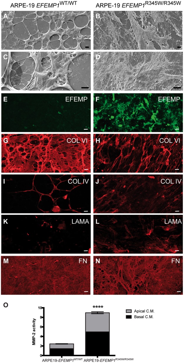
Edited ARPE-19-EFEMP1R345/R345W cells make abnormal ECM. Decellularized transwells of ARPE-19 wild-type (left panels) and ARPE-19-EFEMP1R345/R345W (right panels) cultures after 4 weeks. SEM images show normal ECM (A and C) versus abnormal ECM (B and D) made by wild-type and mutant cells respectively. Immunostaining with antibodies for EFEMP1 (E and F), Col VI (G and H), Col IV (I and J), laminin (K and L) and fibronectin (M and N) show the abnormal structure of the ECM. Scale bars (A–N): 10 µm. (O) Relative MMP-2 activity measured by zymography in apical and basal conditioned media of ARPE-19-EFEMP1WT/WT and ARPE-19-EFEMP1R345/R345W cells (data represented as mean ±SD; t-test; ****P < 0.0001).
