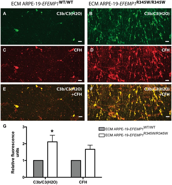
Figure 3.
Abnormal ECM anchors C3b and CFH. Decellularized transwells of ARPE-19 wild-type (left panels) and ARPE-19-EFEMP1R345/R345W (right panels) cultures. Exposed ECM immunostained with antibodies for (A and B) C3b/C3(H2O) and (C and D) CFH. (E and F) Co-staining for C3b/C3(H2O) and CFH. Scale bars 50 µm. (G) Average quantification of C3b/C3(H2O) and CFH fluorescent labeling comparing exposed ECMs (data represented as mean ± SD. ANOVA, *P < 0.05).

