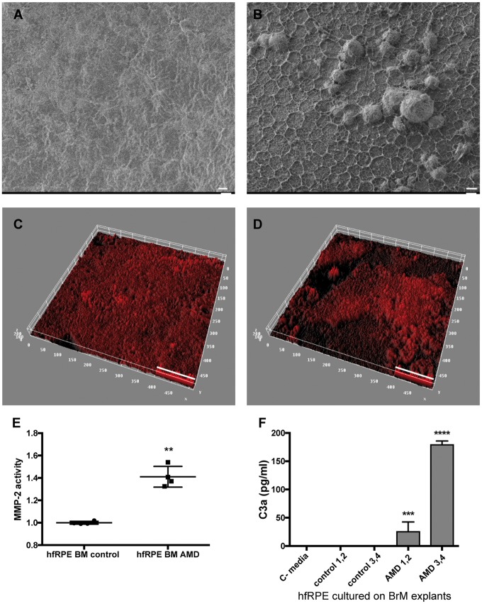
Figure 8.
BrM with AMD alters ECM turnover and causes complement activation by normal hfRPE cells. SEM of BrM from (A) normal and (B) AMD donors. Thick ECM network and drusen-like deposits are visible in the BrM of AMD donors. Representative 3D reconstruction of BrM explants from (C) normal and (D) AMD donors immunostained for collagen IV, and imaged by confocal microscope. Quantification of (C) MMP-2 activity and (D) C3a in conditioned media from hfRPE cells cultured on BrM–choroid–sclera explants from donors without AMD (control 1–4) and with AMD (AMD 1–4). C-media: no cells (data represented as mean ± SD. n = 4 explants/type. ANOVA, ****P < 0.0001, ***P < 0.001, **P < 0.01). Scale bars: (A, B)=10 µm, (C, D)=50 µm.

