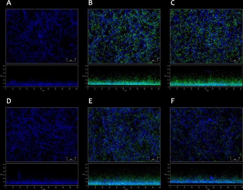
Figure 1.
Formation of mixed biofilms by C. albicans and P. gingivalis wild-type W83 strain (B, E) and P. gingivalis Δppad strain (C, F), observed by confocal laser scanning microscopy. Dual-species biofilms were prepared using 1 × 10⁶C. albicans cells and 1 × 10⁸P. gingivalis cells of each used strain and incubated in RPMI 1640 medium without phenol red for 24 h at 37°C under anaerobic (A–C) or aerobic (D–F) conditions. (A, D) Monospecies fungal biofilms are presented as a reference. Fungal cells (blue) were stained just before imaging with 0.1% Calcofluor White Stain, and bacterial cells (green) were labeled with CFSE prior to addition to the chambers. The images were analyzed with ZEN Lite Microscopy Software.

