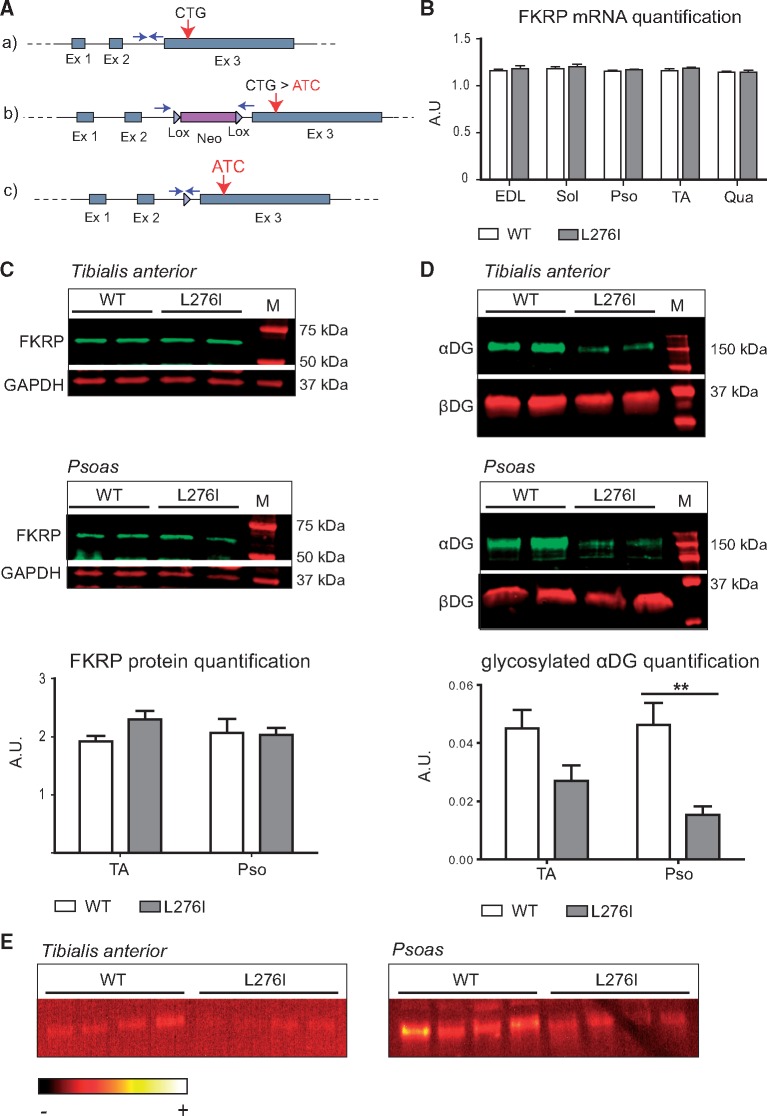
Figure 1.
Generation and molecular characterization of FKRPL276I mouse. A/(A) Representation of the Fkrp locus on mouse chromosome 7 with the position of the L276 codon (CTG) indicated. (B) Plasmid construct used for the homologous recombination. The floxed neomycine resistance cassette was inserted upstream of the third exon. The knock-in mutation (CTG > ATC) was introduced within the third exon. (C) Final recombinant Fkrp allele after deletion of the selection cassette by the CRE recombinase. Arrows represent the primers used for genotyping. (B)/Quantification of Fkrp mRNA by real-time PCR in WT and FKRPL276I muscles normalized by the ubiquitous acidic ribosomal phosphoprotein (P0) mRNA (n = 3; values ± SEM). EDL, soleus (Sol), psoas (Pso), TA and quadriceps (Qua) muscles of 6 months old mice were assayed. AU= arbitrary unit. (C)/Expression of FKRP at protein level by Western-blot on TA and psoas muscles of 2 months old WT and FKRPL276I mice. GAPDH (Glyceraldehyde-3-Phosphate Dehydrogenase) was used as loading control. M= molecular marker. Quantification is shown below the western blots. (D)/Western-blot of glycosylated αDG using IIH6 antibody on TA and psoas muscles of 2 months old WT and FKRPL276I mice. β-dystroglycan (βDG) was used as loading control. M= molecular marker. Quantification is shown below the western blots. (E)/Laminin overlay on TA and psoas muscles of 2 months old WT and FKRPL276I mice. The ImageJ ‘RedHot’ false color scheme was applied to the image.

