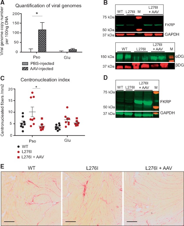
Figure 4.
FKRP gene transfer in FKRPL276I mouse by intravenous administration. (A)/Quantification of viral genome number using qPCR, in psoas (Pso) and gluteus (Glu) muscles of intravenously injected mice, either with PBS (n = 4) or with AAV9-mFkrp (n = 6). (B)/Upper panel: expression of FKRP by western-blot on the psoas muscle of intravenously injected mice, either with PBS or with AAV9-mFkrp. GAPDH is used as loading control. Lower panel: level of glycosylated αDG by western-blot on non-injected and AAV-injected mice in psoas muscle. βDG is used as loading control for αDG. M = molecular marker. (C)/Number of centronucleated fibers per mm2 in psoas (Pso) and gluteus (Glu) of non-injected (n = 8) or intravenously injected with AAV9-mFkrp mice (n = 6). ROUT and Grubbs outlier tests were performed without indicating any outlier. (D)/Expression of FKRP by western-blot on hearts of non-injected and intravenously AAV9-mFkrp injected mice. M = molecular marker. (E)/Sirius red coloration of hearts of non-injected and intravenously AAV9-mFkrp injected mice, showing that even if a high expression level of FKRP is seen in heart after IV injection, it has no consequences on the histological aspect of the tissue. Scale bars = 50 µm.

