Figure 6.
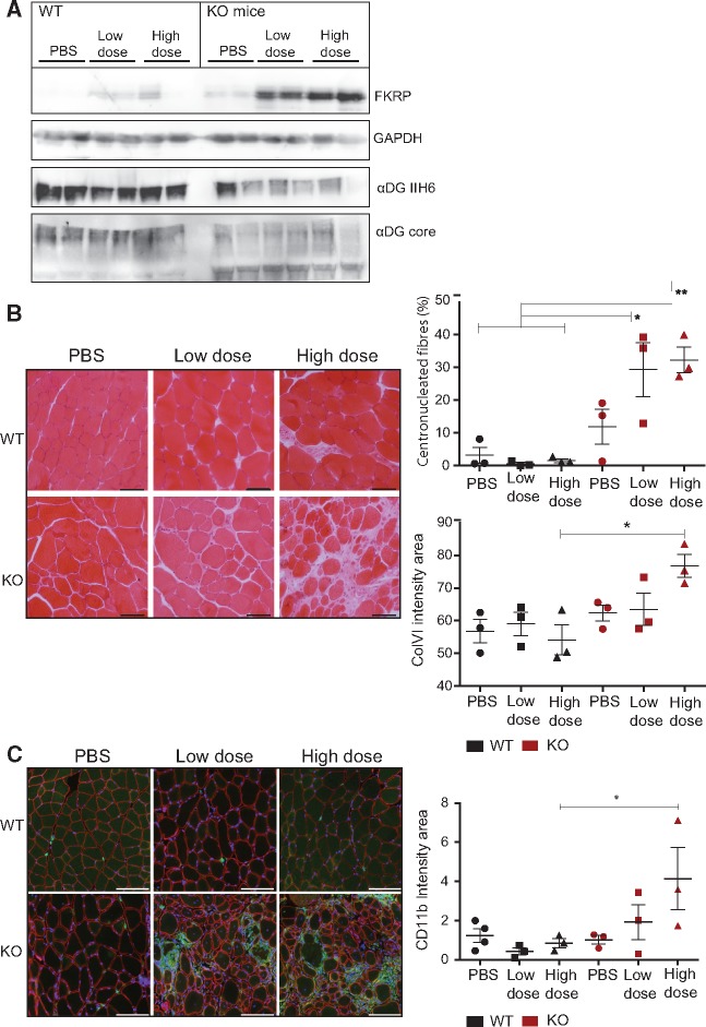
FKRP gene transfer in skeletal muscle-specific Myf5-cre Fktn knockout mice. (A)/Molecular analyses of pooled hindlimb muscle from 8 week-old mice, 4 weeks after intravenous transfer of different doses of AAV9-mFkrp (2 E12 vg/kg and 4 E12 vg/kg). From top to bottom: αDG glycosylation (IIH6 antibody), αDG core protein, FKRP expression (Rbt341) and GAPDH as control. (B)/Left panel: Hematoxine-Eosine staining on histological section of psoas muscle after gene transfer. Scale bars = 100 µm. Right upper graph: percentage of centronucleated fibers in injected mice muscles. Right lower graph: quantification of fibrosis as ColVI intensity (green) of the entire psoas normalized by psoas area (n = 3). (C)/CD11b macrophage marker increased in Myf5-cre Fktn knockout mice 4 weeks after high dose Fkrp gene transfer. Left panel: immunostaining of psoas with CD11b (green), βDG (red) and DAPI nuclear counterstain (blue). Scale bars = 100 µm. Right graph: quantification of CD11b (green, mean fluorescent intensity) of entire psoas normalized by psoas area. (n = 3–4) * P < 0.05. Error bars represent SEM.

