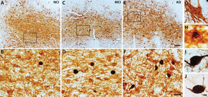
FIGURE 1.
Dual p75NTR/TOC1-immunoreactive neurons. Tissue sections containing the nbM immunostained for p75NTR (G; brown) and TOC1 (J; black) show a modest increase in the number of p75NTR+/TOC1+ neurons during the progression from NCI (A, B), to MCI (C, D) and AD (E, F). Panels (B, D, F) are high-magnification images of boxed area in (A, C, E), respectively. TOC1+ pathology initially appeared within the cytoplasm (G, H) before forming a globose tangle and extending into proximal processes (I). Scale bars: E = 200 μm, and also applies to A and C; F = 50 μm, and also applies to B and D; J = 10 μm, and also applies to G–I.

