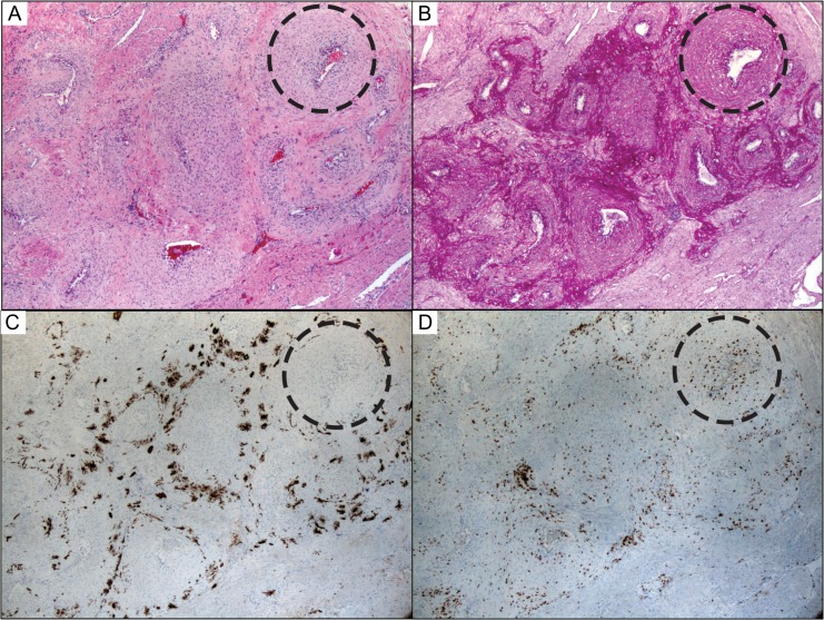
Figure 5.
Radial artery remodeling involves perivascular trophoblasts and accumulation of medial CD3 positive and CD3 negative lymphocytes. (A) Remodeling radial artery at 15 weeks gestation by H&E stain showed medial muscular and extracellular matrix changes appreciated best in complete cross-sections (dashed circle). (B) Periodic acid–Schiff stain highlighted pervivascular adentitia that was invaded by cytokeratin positive EVT cells (C) that do not appear to invade the muscular media. (D) Instead, there are numerous lymphocytes, including CD3-positive T-cells in the walls of these remodeling arteries. Photographs taken using a 5× objective (~50× magnification).

