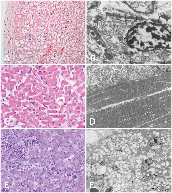
Figure 1.
Pathology of skeletal muscle, heart muscle, and liver tissue. Light microscopy examination using eosin-hematoxylin staining of cardiac muscle at 20x magnification (A), skeletal muscle at 40x (C) and liver at 40x (E). Electron microscopic evaluation of cardiac muscle at 2000x magnification (B), skeletal muscle at 2000x (D), and liver at 1500x (F). Whereas light microscopy did not reveal substantive changes, electron microscopy showed extensive proliferation of mitochondria displacing cytosolic elements in heart, muscle and liver, and mitochondria appear rarefied with poor cristae in all three tissues.

