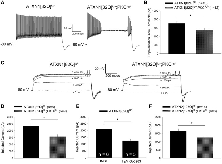
Figure 6.
Increased PKC activity reduces membrane excitability in SCA1 and SCA2 Purkinje neurons. (A) Representative traces from ATXN1[82Q]tg/- and ATXN1[82Q]tg/-; PKCitg/- Purkinje neurons injected with +700 pA of current. ATXN1[82Q]tg/-; PKCitg/- Purkinje neurons undergo depolarization block of repetitive spiking at lower levels of injected current than ATXN1[82Q]tg/- Purkinje neurons, summarized in (B). (C) Representative traces where dendritic calcium spikes were evoked with somatic current injection (injected current amount indicated on the trace) from ATXN1[82Q]tg/- (left) and ATXN1[82Q]tg/-; PKCitg/- (right) Purkinje neurons treated with 1 µm TTX. ATXN1[82Q]tg/-; PKCitg/- Purkinje neurons require less injected current to elicit a dendritic calcium spike than ATXN1[82Q]tg/- Purkinje neurons, summarized in (D). (E) ATXN1[82Q]tg/- Purkinje neurons require less injected current to elicit a dendritic calcium spike in the presence of 1 µM TTX when PKC activity is inhibited with 1 µM Go6983. All slices were pre-incubated with dimethyl sulfoxide (DMSO) or Go6983 for 40 min before recording. (F) ATXN2[127Q]tg/-; PKCitg/- Purkinje neurons require less injected current to elicit a dendritic calcium spike in the presence of 1 µM TTX than ATXN2[127Q]tg/- Purkinje neurons. All data from experiments using ATXN1[82Q]tg/-; PKCitg/- were performed at 20 weeks of age, when dendritic degeneration is detected in these mice. All data from experiments using ATXN2[127Q]tg/-; PKCitg/- were performed at 12 weeks of age, when dendritic degeneration is first detected in ATXN2[127Q]tg/- mice (38). Throughout, data are represented as means with error bars representing S.E.M. * P <0.05. Statistical significance derived by unpaired two-tailed Student’s t-test (B, D, E, F).

