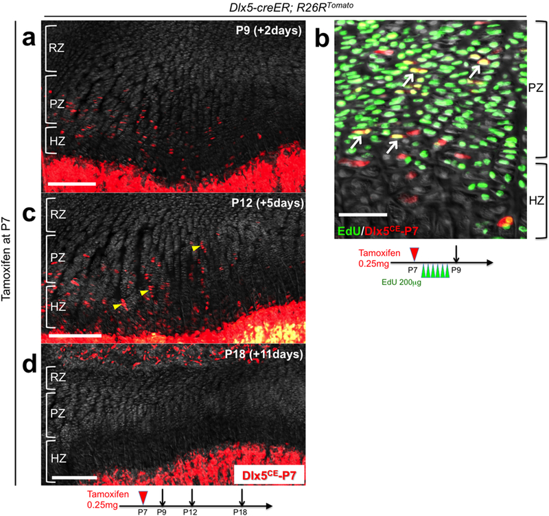
Extended Data Figure 5. Dlx5-creER+ proliferating chondrocytes are not the source of columnar chondrocytes.

(a-d) Cell fate analysis of Dlx5-creER+ proliferating chondrocytes. Dlx5-creER; R26RTomato distal femur growth plates (P7-pulsed). (b): EdU (200μg) was serially injected 6 times at an 8-hour interval between P7 and P9. Arrows: EdU+tdTomato+ cells, arrowheads: short columns (<10 cells). RZ: resting zone, PZ: proliferating zone, HZ: hypertrophic zone. Grey: DAPI and DIC. Scale bars: 200μm (left panels), 50μm (right panel). n=3 mice at each time point.
