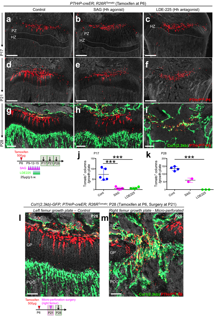
Extended Data Figure 8. PTHrP-creER+ resting chondrocytes form columnar chondrocytes in a Hedgehog-responsive niche-dependent manner.

(a-i) Pharmacological manipulation of Hedgehog signaling. PTHrP-creER; R26RTomato distal femur growth plates (P6-pulsed). Left panels: vehicle control, center panels: SAG (Hh agonist)-treated, right panels: LDE-225 (Hh antagonist)-treated samples. Grey: DAPI and DIC. Scale bars: 200μm. PZ: proliferating zone, HZ: hypertrophic zone. (j,k) Quantification of tdTomato+ columns in PTHrP-creER; R26RTomato distal femur growth plates (P6-pulsed). P17: n=5 (Cont), n=5 (SAG), n=4 (LDE225) mice per group. P28: n=4 (Cont), n=3 (LDE225) mice per group, Data are presented as mean ± S.D. P28: n=2 (SAG). ***p < 0.001, P17 Cont vs. SAG: mean diff. = 67.8, 95% confidence interval [37.5, 98.1], P17 Cont vs. LDE225: mean diff. 66.0, 95% confidence interval [33.9, 98.0], P17 SAG vs. LDE225: mean diff. −1.85, 95% confidence interval [−33.9, 30.2], P28 Cont vs. LDE225: mean diff. 134.5, 95% confidence interval [108.7, 160.3]. One-way ANOVA followed by Tukey’s multiple comparison test. (l,m) Mirco-perforation injury of growth plates. Col1(2.3kb)-GFP; PTHrP-creER; R26RTomato distal femurs (P6-pulsed) at P28. Micro-perforation surgery was performed at P21. (l): left femur growth plate (control), (m): right femur growth plate (micro-perforated). Dotted line: micro-perforated area. SOC: secondary ossification center, GP: growth plate, POC: primary ossification center. grey: DAPI and DIC. Scale bars: 100μm. n=3 mice.
