
Extended Data Figure 10. Absence of tamoxifen-independent recombination in PTHrP-creER line.
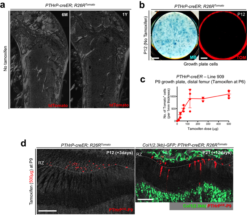
(a) No tamoxifen controls of PTHrP-creER; R26RTomato mice at 6 months (left) and 1 year (right) of age. Red: tdTomato, blue: DAPI, grey: DIC. Scale bars: 500μm. n=3 mice per group.(b) No tamoxifen controls of primary colonies (Passage 0) isolated from PTHrP-creER; R26RTomato mice at P12 without tamoxifen injection. Left panel: Methylene Blue (MB) staining, right panel: red: tdTomato (TOM). Scale bar: 5mm. n=3 mice. (c) Dose-response curve of PTHrP-creER-based recombination. Quantification of tdTomato+ cells in resting zone at P9 in PTHrP-creER (Line909); R26RTomato mice upon a single dose of tamoxifen at P6. x axis: dose of tamoxifen (μg), y axis: the number of tdTomato+ cells per 1 mm thickness. n=3 (0, 31.3, 62.5μg), n=4 (15.6, 125, 250, 500μg) mice per group, data are presented as mean ± S.D. (d) Tamoxifen-induced recombination in P9-pulsed growth plates. PTHrP-creER; R26RTomato distal femur growth plates at P12 (left) and Col1(2.3kb)-GFP; PTHrP-creER; R26RTomato mice at P21 (right). Tamoxifen (500μg) was injected at P9. RZ: resting zone. Green: Col1(2.3kb)-GFP, red: tdTomato, grey: DAPI and DIC. Scale bars: 200μm. n=3 mice.