
Figure 2. PTHrP-creER+ resting chondrocytes are the source of columnar chondrocytes.
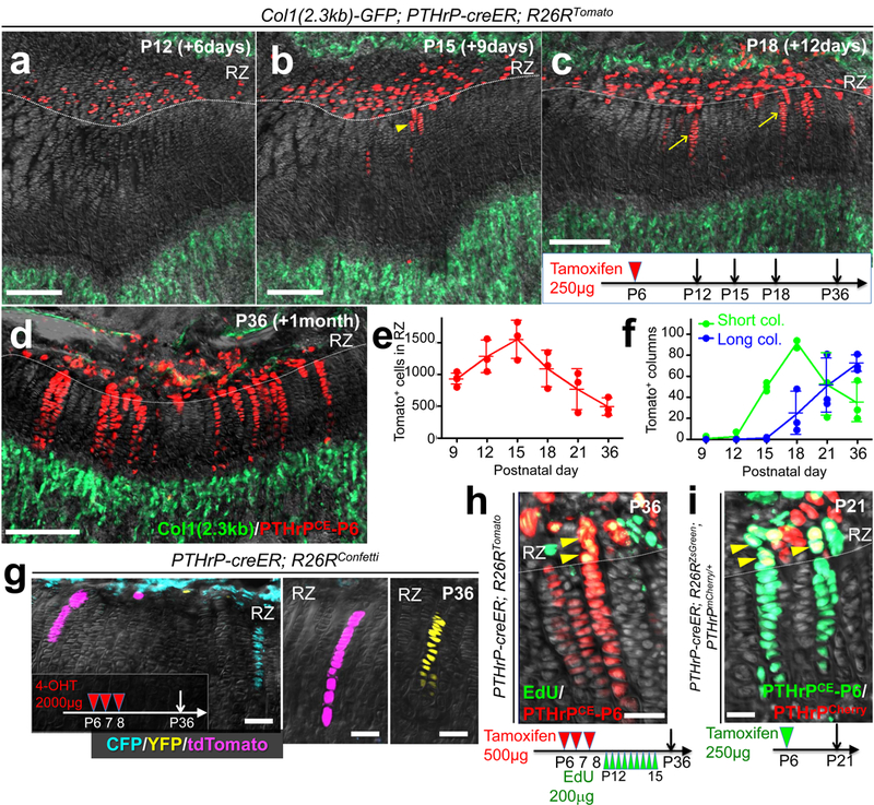
(a-f) Cell fate analysis of PTHrP-creER+ resting chondrocytes. Col1(2.3kb)-GFP; PTHrP-creER; R26RTomato (P6-pulsed) distal femur growth plates. Arrowhead: short column (<10 cells), arrows: long columns (>10 cells). Scale bars: 200μm. (e,f): Quantification of tdTomato+ cells in resting zone (e) (red line) and columns in growth plate (f), short columns (<10 cells, green line) and long columns (>10 cells, blue line). n=5 (P9), n=3 (P12-36) mice per group, data are presented as mean ± S.D. (g) In vivo clonal analysis of PTHrP-creER+ resting chondrocytes. PTHrP-creER; R26RConfetti distal femur growth plates (P6/7/8-pulsed). 4-OHT: 4-hydroxytamoxifen. Scale bars: 50μm. n=3 mice. (h) EdU label-retention assay of PTHrP-creER; R26RTomato distal femur growth plates (P6/7/8-pulsed). Arrowheads: EdU-retaining tdTomato+ cells. Scale bars: 50μm. n=3 mice. (i) PTHrP-mCherry expression in PTHrP-creER; R26RZsGreen; PTHrPmCherry/+ distal femur growth plates (P6-pulsed). Arrowheads: PTHrP-mCherry+ZsGreen+ cells. Scale bars: 20μm. Grey: DAPI and DIC. RZ: resting zone. n=3 mice.