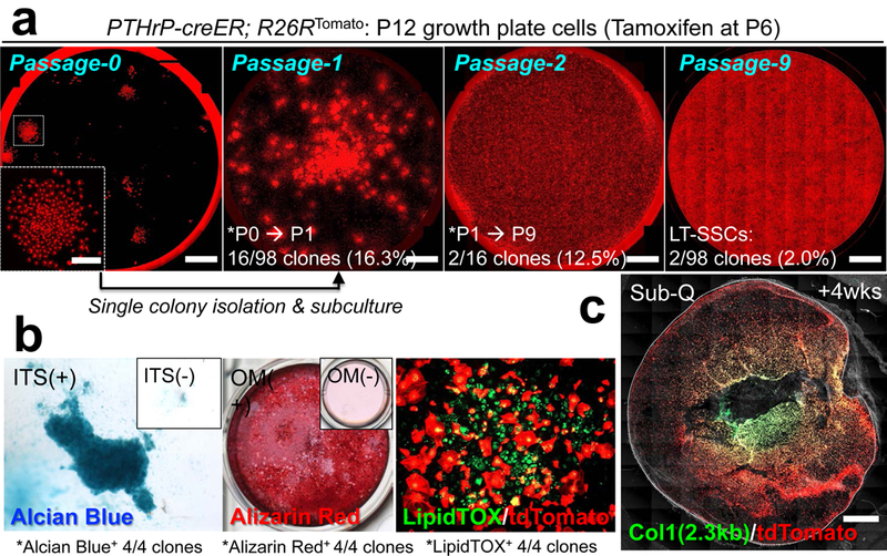
Figure 4. Skeletal stem cell activities of PTHrP-creER+ resting chondrocytes ex vivo.

(a) Colony-forming assay and subsequent passaging of individual PTHrP-creER/tdTomato+ colonies. Inset: magnified view of single colony. Red: tdTomato. Scale bars: 5mm, 1mm (inset). LT-SSCs: long-term skeletal stem cells. n=98 independent experiments. (b) Trilineage differentiation of PTHrP-creER/tdTomato+ clones (Passage 4~7). Chondrogenic (leftmost), osteogenic (left center) and adipogenic (right center) differentiation conditions. Insets: differentiation medium negative controls. ITS: insulin-transferrin-selenium, OM: osteogenic differentiation medium. Four independent clones were tested. (c) Subcutaneous (Sub-Q) transplantation of PTHrP-creER/tdTomato+ clones into immunodeficient mice. Dotted line: contour of the plug. Grey: DIC. Scale bars: 1mm. n=8 mice.
