Figure 5:
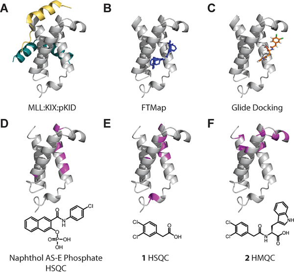
Structural representations of KIX highlighting binding sites mapped onto 1KDX. A) Ternary complex of MLL (gold), KIX (gray), and pKID (teal). B) Select FTMap results displaying probe molecule clusters (blue) in a third site in the protein (PDBID 1KDX). C) Glide docking pose of 2 (orange) using the Schrödinger Maestro Suite. D, E, F) Chemical shift perturbations from 1H-15N HSQC or HMQC experiments. Perturbations greater than one standard deviation are shown in magenta.

