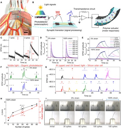
Fig. 4. Organic optoelectronic synapse and neuromuscular electronic system.

(A) Photograph of organic optoelectronic synapse on an internal human structure model. (B) Configuration of organic optoelectronic synapse (photodetector and artificial synapse) and neuromuscular electronic system (artificial synapse, transimpedance circuit, and artificial muscle actuator). (C) EPSCs triggered by single and double visible light spikes (each spike generated presynaptic voltage of −1.1 V for 120 ms). PPF (A2/A1) = 1.42. (D and E) Visible light–triggered EPSC amplitudes of s-ONWST from 0 to 100% strains; (D) SDDP from 120 to 960 ms and (E) SNDP with 1 to 30 spikes. (F) Visible light–triggered EPSC amplitudes of s-ONWST with the International Morse code of “SOS,” which is the most common distress signal. (G) Infrared (IR) and ultraviolet (UV) light–triggered EPSC amplitudes of s-ONWST with the International Morse code of “HELLO UNIVERSE.” (H) Maximum δ of polymer actuator and output voltage generated by s-ONWST according to 0 ≤ nSPIKE ≤ 60 and (I) digital images of the polymer actuator according to 0 ≤ nSPIKE ≤ 100 with 0 or 100% strain.
