Figure 6.

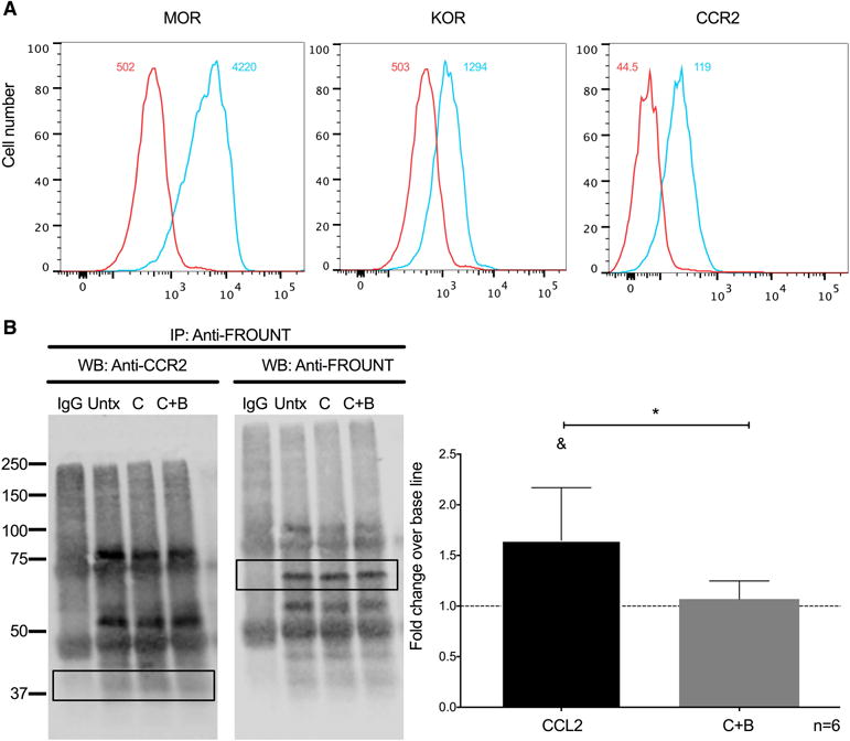
Buprenorphine decreases CCL2-mediated association of FROUNT with CCR2 on THP-1 cells. (A) Expression of MOR, KOR, and CCL2 on THP-1 cells analyzed by flow cytometry. The number depicted in the upper right of each panel is the mean fluorescence intensity for that receptor, and the value in the upper left is the mean fluorescence intensity for the irrelevant isotype matched antibody. (B) THP-1 cells were treated with CCL2 (200 ng/mL) (C), CCL2 plus buprenorphine (40 nM) (C+B), or left untreated (Untx). After 30 seconds of treatment, cells were washed and lysed using digitonin lysis buffer. Immunoprecipitation of FROUNT was performed with 10-15 μg of antibody. Immunoprecipitated proteins were resolved with SDS-PAGE, and analyzed by Western blotting for CCR2 (B; WB: Anti-CCR2; Left panel). As a normalization control, the same membrane was stripped and probed for immunoprecipitated FROUNT (B; WB: Anti-FROUNT; Left panel). Densitrometry was performed on the bands enclosed by the rectangular box in each image. The CCR2 signal was normalized to the FROUNT signal for each condition and then compared to base line. (B, right panel) Results are expressed as mean fold change over base line association of FROUNT to CCR2 (Untx), which was set to one and depicted as a dotted line, ± SD. n = 6 independent experiments. &p<0.05 compared to baseline, *p<0.05 C+B compared to CCL2.
