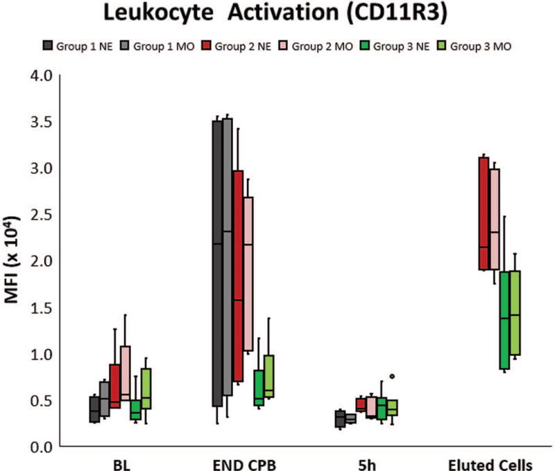
Figure 5.

Leukocyte activation was quantified using flow cytometry by measuring the mean fluorescent intensity (MFI) of surface expression of CD11R3, the porcine analog to human CD11b, on neutrophils (NE) and monocytes (MO) in whole blood. Most pigs in Group 1 and Group 2 had a significant increase in CD11R3 expression at the end of CPB (END CPB) compared to baseline (BL), although individual animal responses varied. The magnitude of this change trended much lower in Group 3. Cells eluted from the L-MOD cartridges post therapy in each of the treated groups had a very high expression of CD11R3, demonstrating that activated leukocytes were sequestered within the device. By 5h post CPB, MFI of circulating cells had returned to the baseline value in all groups, presumably due to extravasation of the activated cells Endpoint of upper whisker, maximum; upper edge of box, third quartile; horizontal line inside box, median; lower edge of box, first quartile; endpoint of lower whisker, minimum; colored dots represent outlier values.
