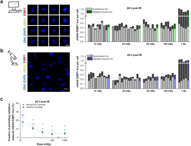
Figure 4.
Inefficient DSB repair after irradiation with low X-ray doses. Non-dividing HOMSF1 cells were irradiated with various doses between 12 mGy and 1 Gy, fixed at 24 h after irradiation and stained for γH2AX, 53BP1 and DAPI. (a,b) Quantification of foci performed by AutoFoci (a) or manually by the experimenter (b). Left panels: For automated foci counting with AutoFoci, single cell images were used, while overview images containing many cells were used for manual foci counting. The scale bars represent 10 µm. Right panels: Each bar represents the mean foci number from 2 duplicate samples irradiated with the indicated dose (dark grey) plotted together with the corresponding mean foci number of unirradiated control samples (light grey). Striped bars indicate that the foci numbers of unirradiated and irradiated cells were similar (sparsely striped) or that irradiated cells showed slightly fewer foci numbers (<0.01 foci per cell) than the corresponding control (densely striped). For automated foci counting at least 5000 cells and for manual counting at least 1000 cells were analyzed per duplicate sample. Foci counting was performed in a blinded manner. The last column for each dose represents the mean value for unirradiated and irradiated cells from the shown 4–12 independent duplicates. Error bars represent the SE. (c) Evaluation of the DSB repair efficiency. The number of the radiation-induced persisting foci as shown in a and b (dark grey part of the columns) was divided by the number of foci induced at 15 min by the corresponding doses by applying an induction rate of 20 foci per cell for 1 Gy. Error bars show the SE from 4–12 duplicates and *indicates a p value < 0.05. Data sets for 12 mGy were tested against 25, 50, 100 and 1000 mGy. The detailed parameters are provided in Materials and Methods.

