
Figure 6.
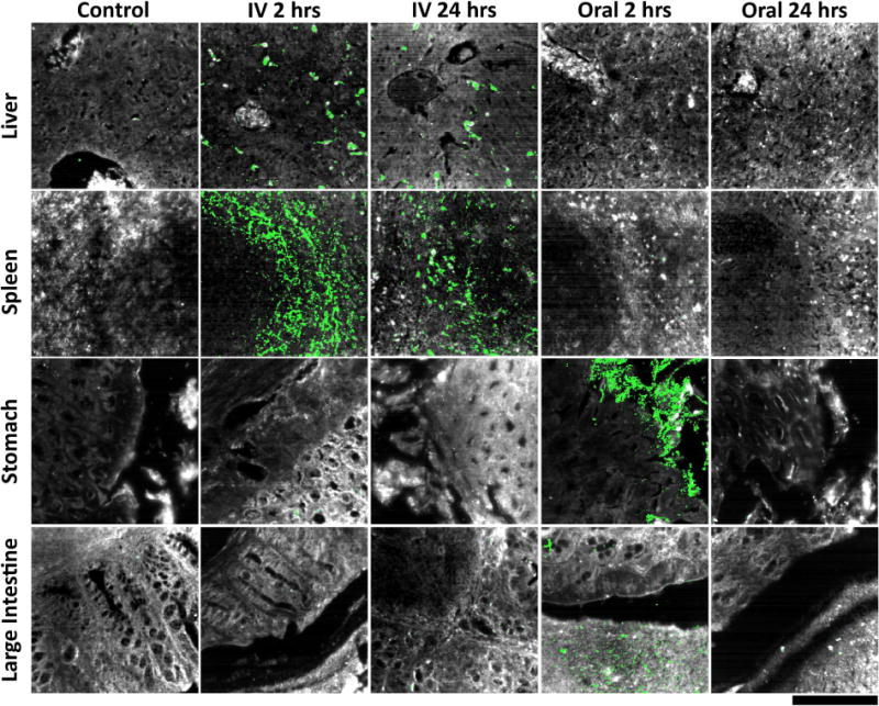
Hyperspectral detection of particle presence in ex vivo tissue sections following intravenous and oral administration. As expected, little to no particle signal was observed in liver, spleen, stomach, and large intestine tissues collected from control mice (far left column). For mice intravenously injected with particles, particle uptake was localized in liver and spleen tissue, while no particle accumulation was apparent within stomach and large intestine tissue 2 hours post-injection (mid-left column). Similar results were observed at 24 hours post-injection, indicating prolonged residence of particles in major clearance organs after intravenous delivery (middle column). By contrast, liver and spleen tissues from mice that received oral particle administration exhibited no particle presence at 2 hours, while stomach and large intestine sections displayed significant particle accumulation (mid-right column). At 24 hours post-oral delivery, particles were not discernible in any of the four tissues analyzed, indicating no translocation to systemic clearance organs as well as virtually complete elimination from the gastrointestinal tract (far-right column). For a quantitative representation of the data please refer to Supplementary Figure S9 within the supporting information. Tissue is depicted in grayscale and particles (identified by their unique spectra) are shown in green for all images presented. Note that these images do not visually convey signal intensity—please refer to Figure S9 for quantitative analysis that does take signal intensity into account. The scale bar at the bottom right represents 100 μm and applies to all images.