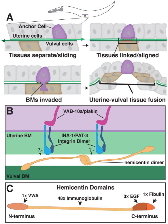
Fig. 1. Anchor Cell invasion in C. elegans and the B-LINK BM-BM adhesion system.
A. A schematic of an L3 stage worm indicating the position of the anchor cell. Top Left – Prior to invasion, the uterine and vulval BMs are separate and the tissues slide along one another. Top Right - The B-LINK complex links the basement membranes (BMs) directly under the anchor cell prior to invasion, aligning the uterine and vulval tissues. Bottom Left – The anchor cell breaches the linked BMs. Bottom Right – The BMs are cleared and the uterine and vulval tissues fuse. B. A magnified inset of the B-LINK complex. Intracellularly, VAB-10A/plakin links the INA-1/PAT-3 integrin heterodimer to the cytoskeleton. Hemicentin is secreted by the anchor cell and may organize into multimers between the BMs that could directly link the BMs. Hemicentin organization is dependent on INA-1/PAT-3 integrin, but it is unknown if this interaction is direct or indirect as indicated by the questions marks. C. Hemicentin is comprised of four major domains: A Von Willebrand Factor type A (VWA) domain, 48 Immunoglobulin repeats, three Epidermal Growth Factor-like (EGF) repeats, and a fibulin family domain.

