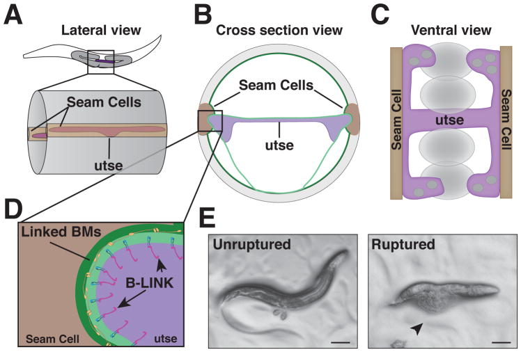
Fig. 4. The utse-hypodermal BM-BM linkage in C. elegans.
A. A mid-body lateral view of an L4 stage worm where the utse and seam cells have linked through their BMs. B. A cross sectional view of the mid body showing the utse connecting to the seam cells on either side of the body. C. A ventral view of the utse-seam cell BM-BM adhesion demonstrating the H-shaped utse and how it acts as a hammock to support the uterine tissue and developing embryos (shown as ovals) during muscle contractions. D. An expanded view of inset in (B) highlighting the B-LINK mediated connection between tissues. E. Left – A wild type worm showing eggs laid as a result of normal egg laying muscle contraction. Right – A worm with a disrupted B-LINK exhibiting external intestine and gonads (arrowhead), which is characteristic of the Rup phenotype as a result of egg laying muscle contraction.

