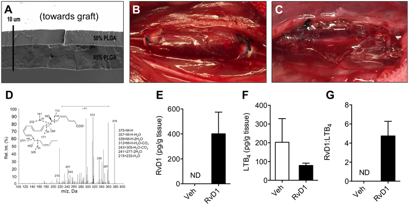
Fig 1.
Perivascular delivery of resolvin D1 (RvD1) to vein grafts in a rabbit carotid bypass model. Poly(lactic-coglycolic acid) (PLGA) films (“wraps”) were constructed using a thin bilayered design, with 1 μg of RvD1 loaded between layers of 50% and 85% PLGA. A, A representative scanning electron microscopy film is shown. B, The thin (<10 μm) design facilitated compliance to the vein graft wall in vivo. C, Alternatively, 1 mg of RvD1 was delivered through a Pluronic gel construct. In vitro studies demonstrated directional drug release from the wrap construct but not from the gels (data not shown). D, Liquid chromatography-tandem mass spectrometry (LC-MS/MS) from vein grafts harvested 3 days after bypass demonstrated delivery of RvD1 to the vessel wall with RvD1-loaded wraps. E, There was no detectable RvD1 at 3 days after bypass with vehicle (Veh) wraps. F and G, There was no detectable RvD1 from vein grafts treated with vehicle and RvD1 gels (n = 1; data not shown). RvD1-loaded wrap treatment resulted in an increase in the ratio of RvD1 to leukotriene B4 (LTB4; n = 3). ND, Not detected.

