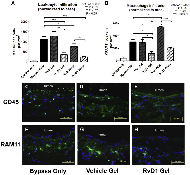
Fig 2.
Perivascular delivery of resolvin D1 (RvD1) decreases inflammatory cell infiltration into vein grafts 3 days after bypass. Jugular veins were harvested and used as ipsilateral carotid interposition grafts in a rabbit model. RvD1 or vehicle (Veh) was delivered to the vein graft at time of bypass through perivascular application of a thin bilayered poly(lactic-co-glycolic acid) (PLGA) wrap or 25% Pluronic F127 gel. Treatment groups consisted of no treatment (bypass only) and perivascular application of vehicle gels, RvD1 gels, vehicle wraps, or RvD1 wraps (n = 3–5). A total of 1 μg of RvD1 was delivered for each drug-loaded group (gel and wrap). Vein grafts were harvested at 3 days after bypass and stained for CD45 and RAM11 to detect leukocyte and macrophage infiltration into the vessel wall. 4’,6-Diamidino-2-phenylindole (DAPI) nuclear counterstaining was also performed. A and B, Quantification of leukocyte and macrophage infiltration was performed using three sections from each graft and normalized to wall area. Inflammatory cells were demonstrated throughout the vessel wall. C-H, Representative images (CD45 and RAM11, green; DAPI, blue). ANOVA, Analysis of variance.

