Figure 5.
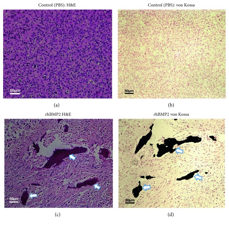
Histological analysis of post‐PET/CT imaging of MDA-MB231 syngeneic tumors: the tumor sections prepared from NBF fixed tissue stained with H&E (a, c) and von Kossa (b, d). Animals injected with BPS as vehicle (a, b) and high doses of rhBMP-2 (15 μg) as IP or direct injection into tumors revealed microcalcifications stained with H&E and von Kossa (c, d). There was nondetectable microcalcification in tumor sections prepared from control tumors (a, b). Magnification = 200x.

