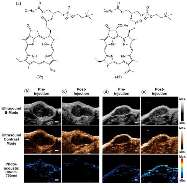
Figure 24.

(a) Molecular structures of the porphyrin–phospholipid conjugates 39 investigated by Lovell et al. (167), Jin et al. (168), Ng et al. (169) and 40 investigated by Huynh et al. (101, 170); (b - e) In vivo imaging in a KB tumor xenograft 10–30 seconds post intravenous injection of bimodal (b, c) and trimodal (d, e) microbubbles derived from 40. US B-mode images show the soft tissue contrast of the tumor, US contrast mode and PA images illustrate the infusion of microbubbles (Scale bar 1 mm). Reproduced with permission from ref. 170. Copyright 2014 American Chemical Society.
