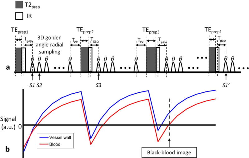
FIG.1.

a: The schematic diagram of the T2 and IR prepared 3D golden angle radial sequence, where the three shown sequence blocks have different duration of T2 preparation. After preparation, spoiled gradient echoes were acquired with flip angle of θ. Tgap is the time between the IR pulse and the first gradient echo, and Tex is the time between the last gradient echo acquisition and the next T2 preparation. S1 and S2 indicate the two adjacent spokes in one shot; S1 and S1′ indicate the two spokes acquired at the same TI in two nearest shots with the same TEprep; S1 and S3 indicate the two spokes acquired at the same TI in two adjacent shots. b: The simulated T2IR signal evolutions of vessel wall and blood. And the black-blood vessel wall image is reconstructed at the near zero point of blood.
