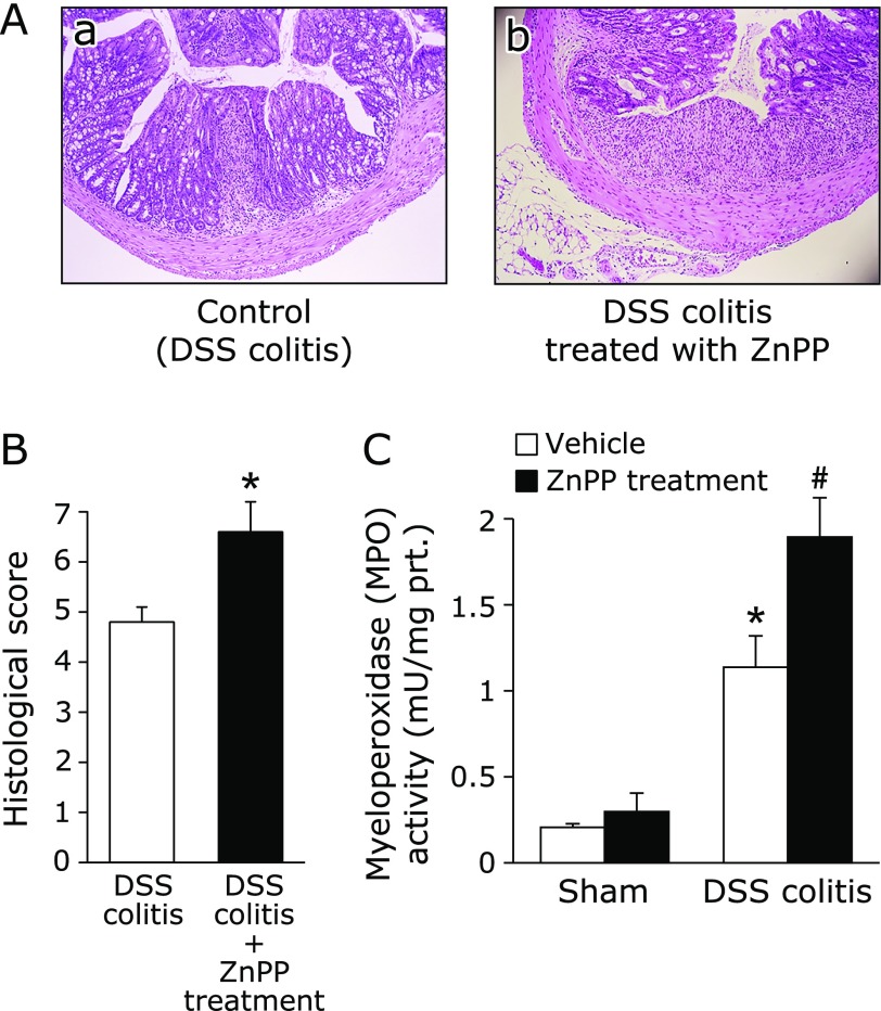
Fig. 3.
Deterioration of colitis by HO-1 inhibition. (A) The histological appearance of colonic tissue in mice with DSS induced-colitis receiving vehicle (a) and those treated with ZnPP (b). Loss and shortening of crypts, mucosal erosions, inflammatory cell infiltration, and goblet cell depletion are seen in (a). In (b), larger erosions are associated with much inflammatory cell infiltration. Hematoxylin and eosin staining. Magnification, ×40. (B) Histological score was evaluated as described in Materials and Methods. The data represent the mean ± SE of 5 mice. *p<0.05 compared with mice with DSS induced-colitis receiving vehicle. (C) Effect of ZnPP on neutrophil accumulation expressed as myeloperoxidase (MPO) activity. Each value indicates the mean ± SE for 7 mice. *p<0.05 compared with mice receiving vehicle, #p<0.05 compared with control mice receiving 2% DSS solution alone.

