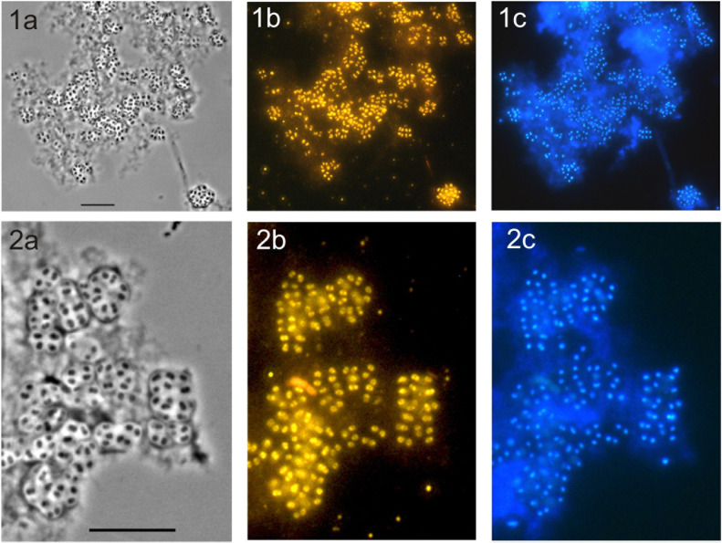
FIGURE 4.
Specific detection of cells of strain SBC82T on micro-particles of amorphous chitin used in growth experiments as a sole source of carbon and nitrogen. The cultures were pictured after 20 days of incubation. Phase-contrast images (a), the respective epifluorescent micrographs of whole-cell hybridizations with Cy3-labeled probe HoAc1402 (b), and DAPI-staining (c) are shown. Rows (1) and (2) demonstrate different fields of view. Enlarged images in row (2) demonstrate characteristic cellular aggregates of strain SBC82T. Bars, 10 μm.

