. 2018 Oct 16;35(11):1140–1155. doi: 10.1039/c8np00037a

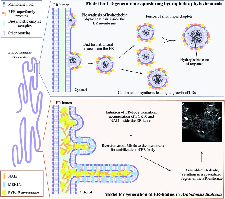
Fig. 5. ER-derived microcompartments, lipid droplets (LDs) and ER-bodies. Top panel: shows the model for lipid droplets sequestering hydrophobic phytochemicals inspired by Laibach et al. 2015, Journal of Biotechnology.160 During biosynthesis, the hydrophobic phytochemical such as natural rubber accumulate inside the ER membrane leaflet causing bud formation and release of LD confined by an ER membrane monolayer. Small LDs may fuse to larger LDs at the same time as continued biosynthesis may occur resulting in growth of LDs. Lower panel: illustrates the development of ER-bodies in Arabidopsis thaliana in which the myrosinase PYK10 accumulates inside the ER lumen together with the soluble protein NAI2. This results in bud formation and triggers the recruitment of the membrane proteins MEB1 and MEB2 that interacts with NAI2, elongating the bud into the rod-shaped ER-body (1 μm× 10 μm) highly enriched with PYK10 and other β-glucosidases. Confocal image of ER-localized GFP (grey ER-network) in ER-bodies (white dilated ER cisternae) in Arabidopsis thaliana is adapted from Nakano et al. 2014, Frontiers in Plant Science.150.

Fig. 5