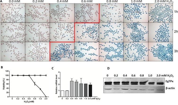
Figure 1.
Viability and EgTPx expression of protoscoleces in the presence of H2O2. (A) Methylene blue staining of protoscoleces exposed to different H2O2 concentrations for 1 h, 2 h and 3 h. (B) Survival of protoscoleces incubated in medium with different concentration of H2O2 for 1 h (triangles). Protoscoleces incubated in medium without H2O2 were used as a control (open boxes), n ≈ 200. (C) Quantitative analysis of mRNA transcripts for expression of EgTPx. (D) Western blotting showing expression of EgTPx in different concentrations of H2O2. The bars indicate mean ± S.E.M.

