Figure 1.
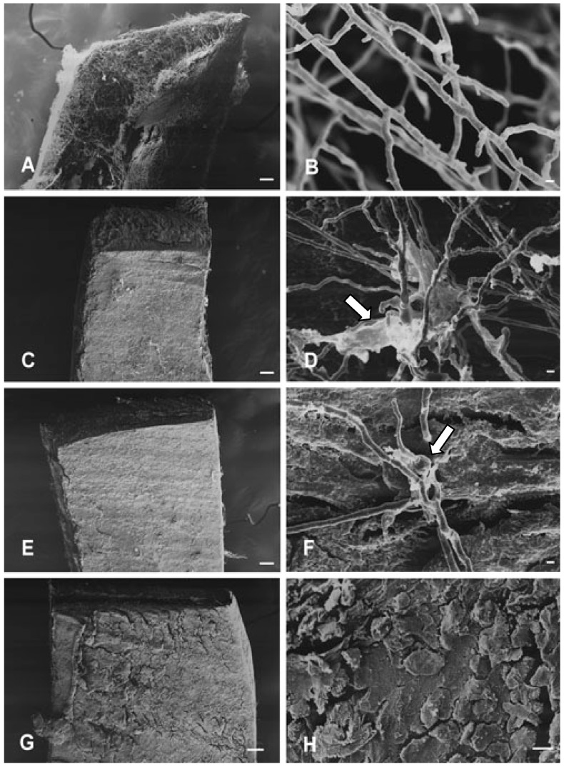
Scanning-electron microscopy showing mycelial structures of T. mentagrophytes cultured on nail fragments for 72 h, at 28 °C: (A-B) Control, (C-D) Treatment with TIO C 55 μmol L-1. (E-F) Treatment with TIO C 110 μmol L-1. (G-H) Treatment with TIO C 220 μmol L-1. View of nail fragment in (A) colonised by T. mentagrophytes. (C) and (E) show the inhibitory effect of TIO C on the invasion of nails. In (D) and (F) arrows indicate excretion of fibrillar materials and swollen structure. Figures (G) and (H) show that no mycelial growth can be seen on nail scales. Bars: 100 μm (A,C,E,G); 10 μm (H); 1 μm (B,D,F).

