Figure 8.

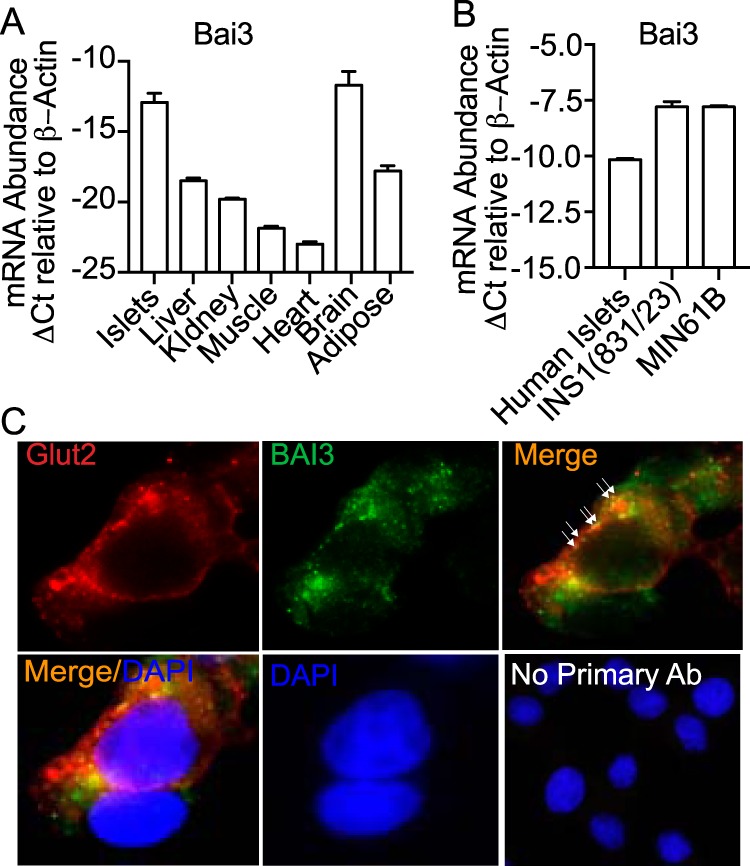
Tissue expression of the BAI3 receptor. A, tissues were isolated from 10-week-old C57BL6/J mice and total mRNA was harvested from islets, liver, adipose, gastrocnemius (gastroc), kidney, heart, and brain. B, human islets, INS1(832/13), and MIN6B1 cells were harvested and total mRNA was prepared. Quantitative real-time PCR was performed to determine the relative mRNA abundance of BAI3. The data were normalized to the β-actin gene. The ΔCt was calculated by subtracting the raw Ct of the BAI3 gene from the raw Ct of the β-actin gene. Values are mean ± S.E. of n ≥ 3. C, immunofluorescence by confocal imaging was performed in INS1(832/13) β-cells using anti-BAI3 antibody (green). The Glut 2 (red) antibody was used as a plasma membrane marker. The localization of BAI3 on the plasma membrane is shown by arrows or as colocalization as a merge in orange. No primary antibody control was used to show that no nonspecific signal was observed from the secondary antibody (scale bar = 10 μm).
