Figure 7.

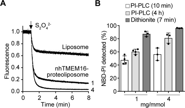
nhTMEM16-mediated scrambling of NBD-PI probed by dithionite and PI-PLC. A, liposomes and nhTMEM16 proteoliposomes at a PPR of ∼1 or 4 mg/mmol containing trace amounts of NBD-PI (Fig. S2) were reconstituted as in Fig. 4B, except for the addition of 250 μm Ca2+ in the reconstitution solution. Dithionite was added after 1 min, and fluorescence was recorded for 8 min. Traces of liposomes and nhTMEM16 proteoliposomes (PPR of ∼1 or ∼4 mg/mmol, indicated next to the corresponding trace) are representative of three independent experiments. B, nhTMEM16 proteoliposomes at PPRs of ∼1 and ∼4 mg/mmol containing trace amounts of NBD-PI were prepared as above in the presence of 250 μm Ca2+. The extent of PI-PLC–mediated hydrolysis of NBD-PI after 10 min (white bars) and 4 h (light gray bars) was determined as described under “Experimental procedures,” and reduction of NBD-PI fluorescence by dithionite (dark gray bars) was determined as described above. The data represent mean values ± standard deviations from three or four measurements.
