
Figure 2.
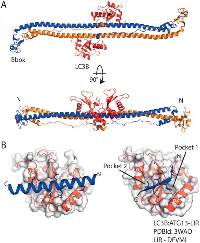
The coiled coil of Trim5α binds LC3B through a helical motif. A, cartoon representation of the RhT5 88–296 EK/RD–LC3B protein complex. Chain A/B (blue/orange) form the Trim5α antiparallel coiled coil dimer, LC3B molecules are red, and zinc atoms are shown as spheres (silver). B, expanded view of the LC3B-binding site (left) and comparison with a typical β-strand LIR motif (LC3B–ATG13-LIR PDB code 3WAO). Both the helical and β-strand LIR occupy and proceed in the same orientation through the LC3B-binding site.