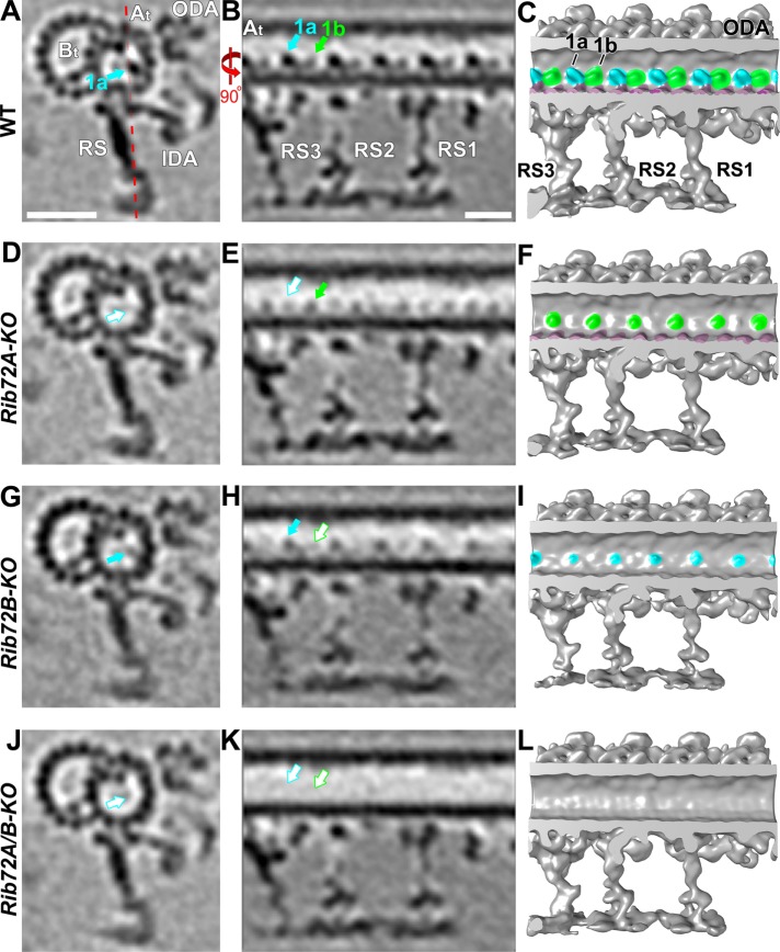
FIGURE 5:
Losses of RIB72A and RIB72B affect the MIP1 structure differently. (A–L) Averaged tomographic slices (A, B, D, E, G, H, J, K) and isosurface renderings (C, F, I, L) in cross-sectional (A, D, G; from proximal) and longitudinal views (B, C, E, F, H, I; proximal on the right) of Tetrahymena axonemal repeat units of RIB72A-KO (D-F), RIB72B-KO (G–I), and the double mutant (J–L) showed different defects in the MIP1a (blue) and/or MIP1b (green) structure compared with WT (A–C). The RIB72A-KO mutant is missing MIP1a (open blue arrow) but not MIP1b (solid green arrow); the RIB72B-KO mutant lacks MIP1b (open green arrow) but not MIP1a (solid blue arrow). The double KO shows both defects, that is, MIP1a and MIP1b are missing (open blue and green arrows). Red dotted line in A indicates slice position of longitudinal tomographic slices, for example, in B. Other labels: At, A-tubule; Bt, B-tubule; IDA, inner dynein arms; ODA, outer dynein arms; RS, radial spokes. Scale bars: 25 nm (A, valid also for D, G), 16 nm (B, valid also for E, H).

