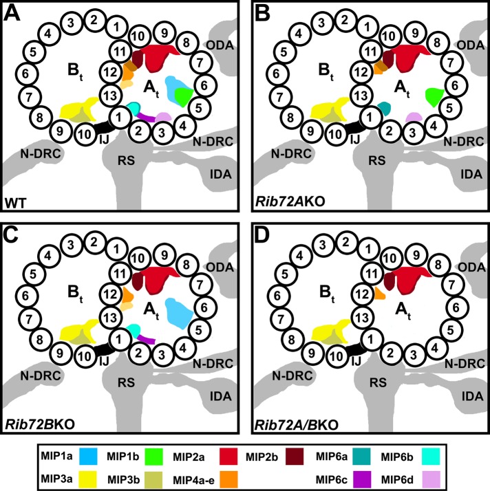
FIGURE 9:
Summary schematics of MIP locations in the DMTs of WT (A), RIB72A-KO (B), RIB72B-KO (C), and RIB72A/B-KO (D) cilia. The structural comparison between the averaged DMTs of WT and mutant axonemes revealed that loss of RIB72A causes the loss of MIPs 1a (blue) 4b (light orange), 4d (yellow), 4e (tomato) (partially missing), 6b (aqua), and 6c (purple). The absence of RIB72B results in the loss of MIPs 1b (green), 4e (tomato), 6a (teal), and 6d (mauve). The defects in the double knockouts are additive. Coloring of all MIPs is shown in the color legend. Other labels: At, A-tubule; Bt, B-tubule; IDA, inner dynein arms; IJ, inner junction; N-DRC, nexin dynein regulatory complex; ODA, outer dynein arms; RS, radial spokes; 1–13, PF numbers.

