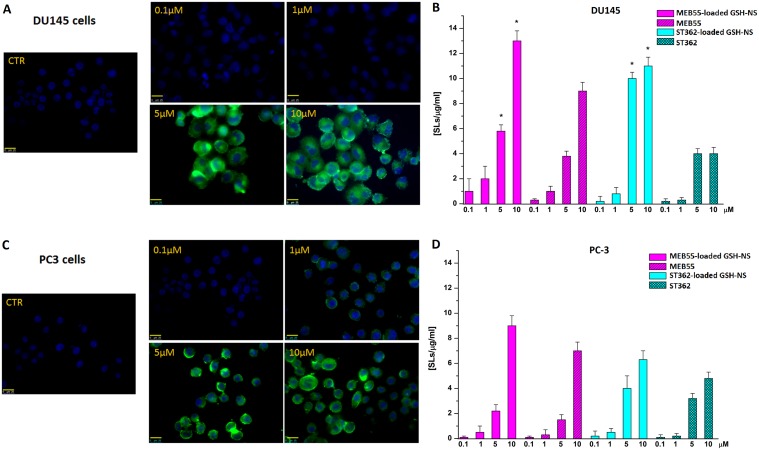
Figure 11.
Fluorescence images (400X magnification) of the GSH/pH-NS (green) in DU-145 (A) and PC-3 (C) cells. Comparison between untreated cells (CTR) and cells treated with 0.1–10 μM GSH/pH-NS after 4 h; (B) Intracellular SL content in DU-145 (B) and PC-3 (D) cells 24 h after treatment with 0.1–10 μM free SLs or SL-loaded GSH/pH-NS.

