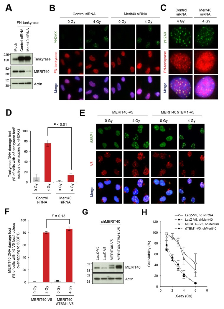
Figure 3. Disruption of MERIT40-mediated recruitment of tankyrase to DSBs decreases the viability of X-ray-irradiated A549 cells.
(A) Western blot of A549 cells with ectopically expressed and nuclear localized tankyrase and depletion of MERIT40. Cells were transfected with an expression vector for FN-tankyrase and MERIT40 siRNA as indicated. After 48 h, western blot analysis was performed with the indicated antibodies. β-actin served as loading control. (B) Indirect immunofluorescence staining. Cells treated as in A were irradiated with 4 Gy of X-ray, incubated for 6 h, and subjected to immunofluorescence staining with anti-γH2AX (green) and anti-tankyrase (red) antibodies. Nuclei were counterstained with DAPI (blue). (C) Magnified views of representative cells in B. (D) The percentages of the cells with more than five tankyrase foci/nucleus that overlapped with γH2AX foci shown in B and C were quantified. Statistical analysis was performed by student's two-tailed t-test. (E) Foci formation of MEIT40ΔTBM1 mutant. V5-tagged, siRNA-resistant MERIT40 (wild-type and ΔTBM1 mutant) was stably expressed in A549 cells. These cells were transfected with MERIT40 siRNA to deplete only endogenous MERIT40. After 48 h, cells were irradiated with 4 Gy of X-ray, followed by incubation for 6 h. Cells were subjected to immunofluorescence staining with anti-53BP1 (green) and anti-V5 (red) antibodies. Nuclei were counterstained with DAPI (blue). (F) The percentages of the cells with more than five V5 foci/nucleus that overlapped with 53BP1 foci in E were quantified. Statistical analysis was performed by Student's two-tailed t-test. (G) Western blot analysis of A549 cells that stably expressed MERIT40 shRNA and either MERIT40-V5 or MERIT40ΔTBM1-V5. Cells, in which shRNA-resistant V5-tagged MERIT40 (wild-type or ΔTBM1 mutant) or LacZ was stably expressed, were infected with lentivirus for MERIT40 shRNA. After selection, cell lysates were prepared and subjected to western blot analysis with the indicated antibodies. β-actin served as loading control. (H) X-ray sensitivity of the cells in (G) Cells were irradiated by indicated doses of X-ray. After a 10-day incubation, the numbers of colonies were quantified. Data were normalized with the colony numbers at 0 Gy, in which cell viability was defined as 100%. Three independent experiments were performed and each experiment was performed in triplicate.

