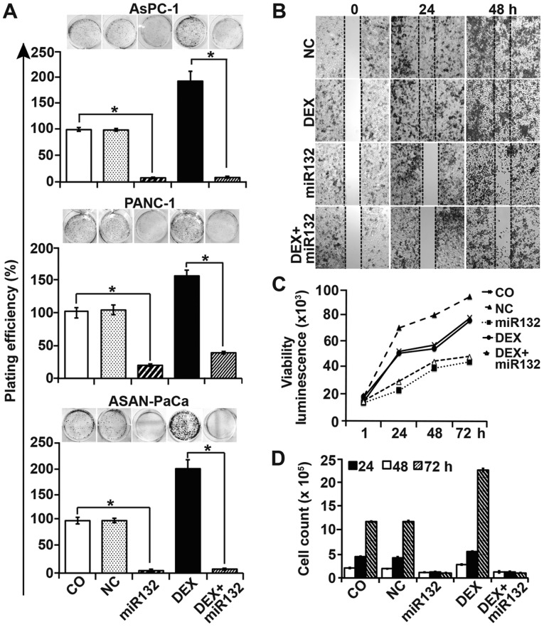
Figure 4.
Cancer progression features are inhibited by miR-132. (A) AsPC-1, PANC-1 and ASAN-PaCa cells were transfected with 50 nM miR-132 mimics or a negative miR control (NC). At 8 h later, the cells were treated with 1 µM DEX in the presence or absence of miR-132. After 48 h, the cells were resuspended in complete medium and plated at a low clonogenic density in 6-well tissue culture plates. After 14 days, colony-forming assays were performed and evaluated as described in the materials and methods. (B) PANC-1 cells were transfected as aforementioned. At 48 h after transfection, the cells were seeded at a high density in ibidi culture insert 24-well plates. After 24 h, once the cells had attached and reached ~90% confluency, the inserts were removed to leave a defined 500-µm-thick scratch. Images of the cell-free gap were obtained immediately (0 h), and at 24 and 48 h after removal of the inserts. (C) PANC-1 cells were treated as afore-described, and cell viability was measured using a RealTime-Glo™ MT Cell Viability Assay. (D) The number of PANC-1 cells was determined by the use of a Coulter counter after 24, 48 and 72 h of treatment. Data are presented as the means ± SD. *P<0.05. DEX, dexamethasone; NC, negative miR control.

